Pictures of Cerebral Aneurysms
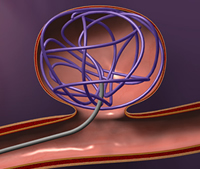
Treatment of a Brain Aneurysm with Detachable Platinum Coils
Tiny platinum coils are threaded through a microcatheter and pushed into the aneurysm. The coils are flexible enough to conform to the aneurysm shape.

The aneurysm is filled in with coils, obstructing the flow of blood into the aneurysm. Each coil is attached to a delivery wire, allowing the physician to reposition or withdraw the coil to ensure ideal placement. Once properly positioned within the aneurysm, the coil is detached from the delivery wire using an electrolytic detachment process (electrical charge).
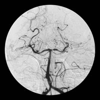
Angiograms of Aneurysms Pre and Post Treatment
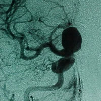
Angiogram of an aneurysm before treatment. The aneurysm is the dark bulge on the vessel.
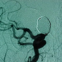
Angiogram of an aneurysm after endovascular coiling treatment. The aneurysm has been filled in with coils, so blood can no longer flow into the aneurysm. The aneurysm now appears as a silver bulge on the angiogram.
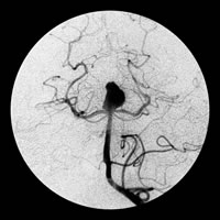
Angiogram of an aneurysm before treatment. The aneurysm is the dark bulge on the vessel.
Angiogram of an aneurysm after endovascular coiling treatment. The aneurysm has been filled in with coils, so blood can no longer flow into the aneurysm. The aneurysm now appears as a silver bulge on the angiogram.
3D Angiography of Aneurysms
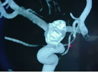
3-D angiography picture of a brain aneurysm. The aneurysm is the bulge on the vessel in the middle of the picture. 3-D angiography is used to determine the dimensions and shape of aneurysms.